VisiView® Software
Flexible and microscope platform independent software
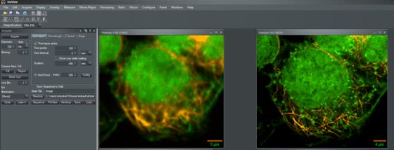
VisiView® is a high performance imaging software from Visitron Systems GmbH for BioImaging applications. It is specially designed to meet the needs for high speed image acquisition and processing with ease of use. Our software controls complex automated microscopes and microscope equipment in combination with multidimensional acquisition and analysis. Its multitasking ability supports simultaneous image acquisition and analysis. The VisiView® software represents the philosophy of simple operation and seamless integration of applied standards.
VisiView® Options
VisiSRRF Super Resolution Algorithm
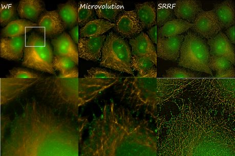
What is SRRF?
SRRF is basically an algorithm which identifies radial fluctuations in a series of images and recalculate the origin of fluorophores by the degree of local radiality and its temporal correlation (Gustafs-son et al. 2016, Culley et al. 2018). The resolution which can be achieved with SRRF is around 70nm. The idea was developed in the lab of Ricardo Henriques and ended up in an GPU-enabled ImageJ Plugin called „NanoJ-SRRF“ (https://henriqueslab.github.io/resources/NanoJ-SRRF/).
AI - Artifical Intelligence Options
Deep Learning Approaches in Microscopy with VisiView®
Deep learning using convolutional neural networks in microscopy applications opens up a new dimension in pattern recognition, image enhancement and object segmentation where classical methods come to their limits. We are excited to share this new VisiView feature with scientists in life science research. Even where the human eye reaches its detection limits, the AI technology outperforms the traditional image analysis by improving resolution and segmentation.
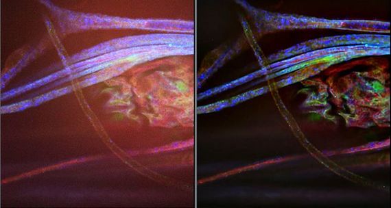
Realtime 2D-Deconvolution
Populare widefield microscopy is a powerful optical method to visualize cellular and molecular processes involved in cells. Although fluorescent blurring from out-of-focus light is an limiting factor, but image deconvolution algorithms can reverse this out-of-focus light artifact. The 2D realtime deconvolution technology, helps to improve cellular image’s resolution and contrast by mathematical deblurring of the image. As a result, images and fine detail become sharper while maintaining quantitative information.
Realtime 3D-Deconvolution
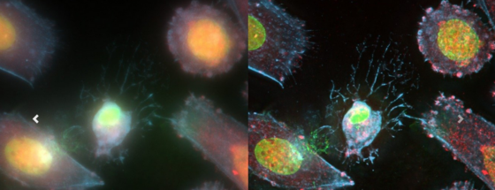
The Microvolution® software delivers nearly instantaneous deconvolution by combining intelligent software programming with the power of a GPU.Confocal microscopy is an oft-used technique in biology. Deconvolution of 3D images reduces blurring from out-of-focus light and enables quantitative analyses, but existing software for deconvolution is slow and expensive. We present a parallelized software method that runs about 200 times faster than other software by running on a low-cost graphics processor board (GPU).
VisiView® Automated Shading and Background Correction
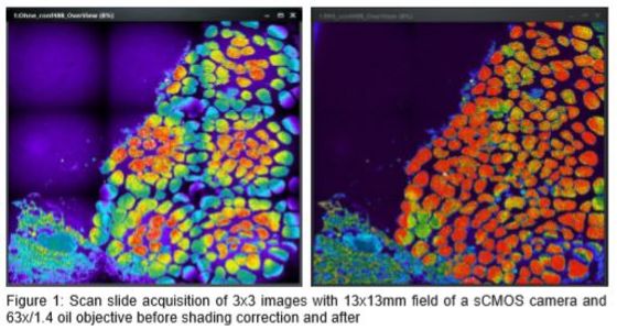
Shading ruins all our microscope images quality. As a consequence the intensity of objects can’t be reliably compared across a single or multiple images. Also you won’t publish images showing ugly holes in the signal. There are two main causes of shading in microscopy, number one is uneven illumination, number two is dirt.