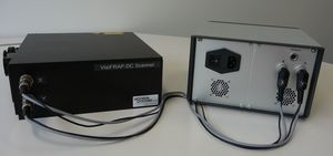

2D-VisiFRAP-DC System with integrated laser
Full Range Photo-Manipulation Imaging System
New: - from 355nm to VIS Laser – NO limit with laser lines
- With unlimited number and size of regions
- Auto-calibration
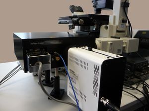
The latest model extension of the Visitron System 2D-FRAP scanner family convinces by its compactness and flexibility. The new design allows the direct coupling of up to two lasers in the Galvo-scan-head without laser fibres. An additional laser fibre input offers the combination of the system with other VIS-laser lines if required.
This design allows the easy combination of UV laser (355nm / 405nm) used e.g. in ablation experiments, together with VIS laser for FRAP or other photomanipulations of the specimen. This unique new scan head opens new dimension in biological studies of living cells.
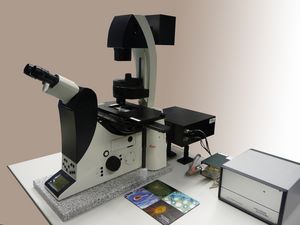
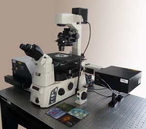
The 2D-VisiFRAP-DC System is based on a pair of galvanometer-driven scan mirrors directly computer controlled by the VisiView imaging software. The Scan head is mounted and coupled to the FL-excitation port of the microscope, where it directly manipulate the specimen.
FRAP on the fly
The optimized system components allow simultaneous FRAP and imaging at single mouse
click on any position in the live image. This new feature in the VisiView FRAP software is
minimising any loss of temporal information and by taking advantage of the flexibility and high
speed positioning of the VS-FRAP scanner. The unique “FRAP on the fly” solves perfectly the
major demand for photo-manipulation experiments.
Auto-Calibration
With the automatic signal and spot detection of our VisiView imaging software, the autocalibration
algorithm calibrates the FRAP scanner. It shows the laser spot and the accuracy of
the calibration in several regions on the display. This tool makes it easy to use different
objectives and filters. The high reproducibility of the system settings saves time and improves
your work.
Typical Application:
- Ablation studies
- Cell membrane analysis
- Monitoring of surface trafficking
- Nucleocytoplasmic shuttling
- Protein diffusion studies
- Region-specific Photoactivation
- Acceptor photobleaching
- Photoconversion studies